Root canal treatment is a complex process that requires great attention, precision and the ability to work with the finest instruments. The main stages:

- Diagnosis: determining the condition of the tooth and the disease. For this purpose, different types of images can be used, such as computed tomography (CT) or dental X-rays.
- Preparation: the patient receives anaesthesia to ensure comfort during the procedure.
- Opening the tooth cavity: the doctor opens access to the pulp chamber of the tooth, where the neurovascular bundle is located.
- Detailing: determining the entrances to the root canals and measuring their length.
- Instrumental treatment: special sets of instruments are used to create a conical shape of the canals and clean them. This allows us to prepare the canals for further antiseptic treatment.
- Drug treatment: drugs are used to maximise the cleaning of the canals from bacteria and prevent their reproduction. Ultrasonic instruments can be used to increase the effectiveness of the antiseptic treatment.
- Filling of the canals: it is possible to fill them with a temporary material that enhances the anti-inflammatory effect, and after a while - permanent obturation.
- Restoring the appearance of the tooth: the method and material depends on the degree of destruction. It can be a filling, restoration, inlay, or crown.

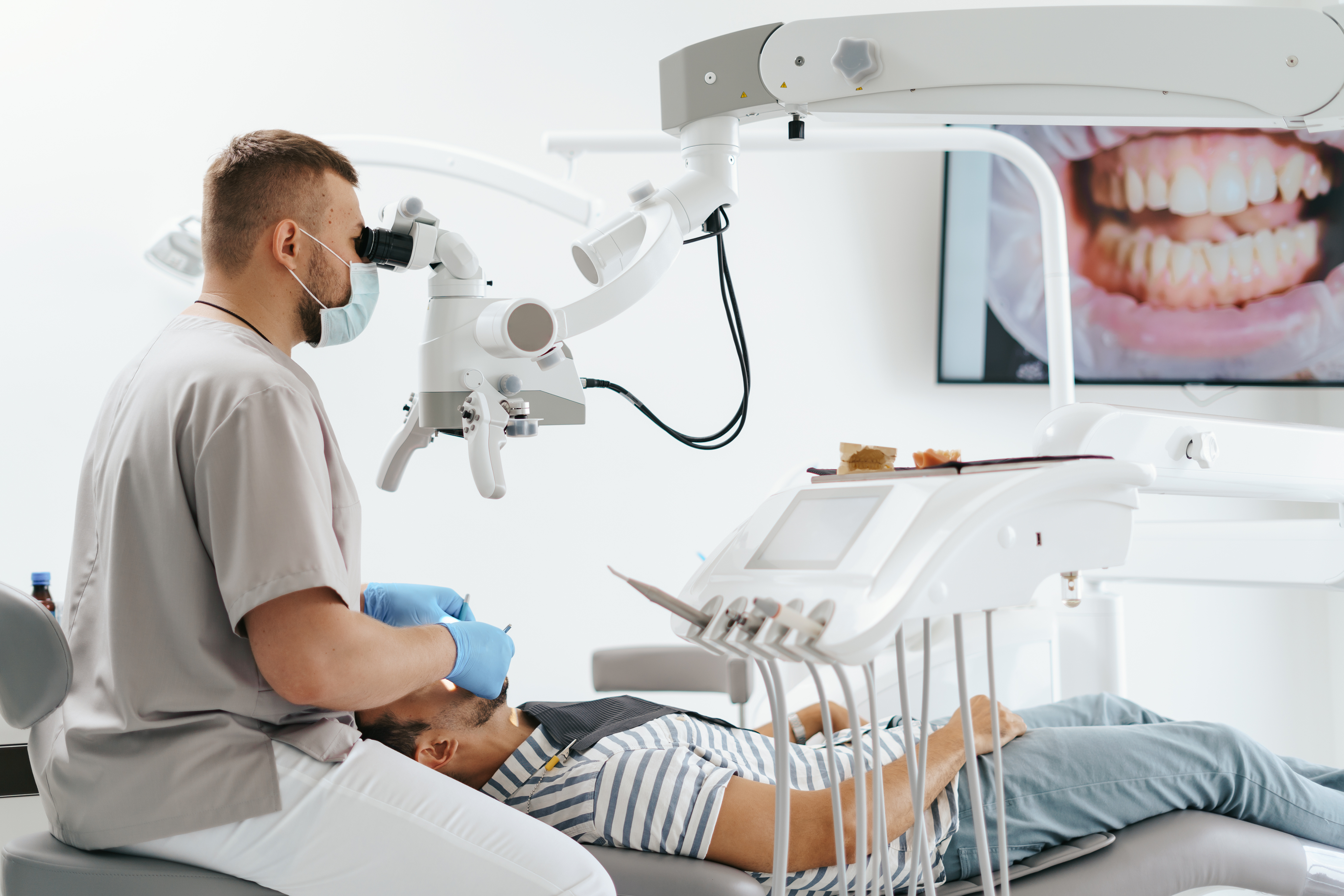
Root canal treatment under a microscope is one of the most advanced methods of modern endodontics used to achieve the most accurate and effective results. Here are some key advantages of this approach:
- Increased precision: the use of a microscope allows for a detailed image of the root canals, which greatly facilitates their treatment. The risk of complications due to improper access to the canals or incomplete cleaning is reduced.
- Process control: the microscope allows the doctor to visually control every stage of treatment, from the search for root canals to their filling. This helps to avoid mistakes and ensure high-quality treatment.
- Optimal use of instruments: the microscope allows you to control the instruments with great precision, which is especially important when preparing root canals. This ensures more precise and efficient canal treatment, which affects the final result.
- Evaluation of results: thanks to the microscope, the doctor can assess the cleanliness of the canal walls, detect possible remnants of tissue or filling materials, which allows them to be removed in time and ensure high-quality canal filling.